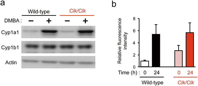
Figure 2.
Induction of Cyp proteins and formation of DNA adducts in the skin of wild-type and Clk/Clk mice after the DMBA treatment. (a) Protein levels of Cyp1a1 and Cyp1b1 in the skin of wild-type and Clk/Clk mice after the DMBA treatment. Skin homogenates were prepared 12 hours after the treatment with 100 μg DMBA. Plus and minus indicate the treatment with DMBA and vehicle (200 μL acetone), respectively. Full-size images are presented in Supplementary Fig. 3. Western blotting data were confirmed in more than three mice in each group. (b) Amounts of DMBA-DNA adducts in the skin of wild-type and Clk/Clk mice. DMBA was applied twice at 8-hour intervals. DNA was extracted from the skin of mice at 0 or 24 hours after the second DMBA treatment. The fluorescence intensity derived from DMBA-DNA adducts was measured, and intensity was normalized by DNA contents in samples (Means ± s.e.m.; n = 3–4).

