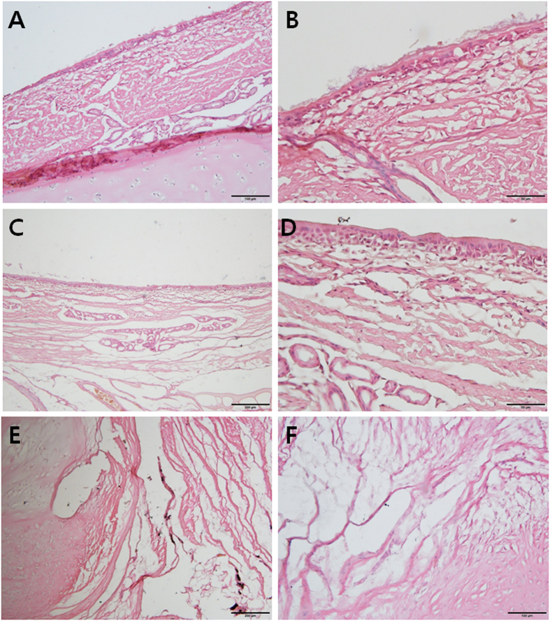
Figure 5.
Hematoxylin and eosin staining of the trachea of pig no. 4, harvested 84 days after the surgery. (A) Mucociliary epithelium was noted on the fibrous connective tissue in the normal tracheal lumen. (B) Mucociliary epithelium in the normal tracheal lumen was revealed at high magnification (×400). Pseudostratified columnar epithelium with cilia was evident. (C) Epithelial cells with scanty cilia were noted on the muscular fiber tissue in the neo-tracheal lumen. (D) The surface of the neo-trachea was evaluated at high magnification (×400). Stratified squamous epithelium without cilia was observed. (E) A transition zone between the normal trachea and the neo-trachea was evident. The cartilaginous tissue (left portion) was faced with muscular fibrous tissue (right portion). (F) A transition zone between the muscular fibrous tissues of the neo-trachea and the cartilaginous tissue of the normal trachea was noted at high magnification (×200).

