Figure 6.
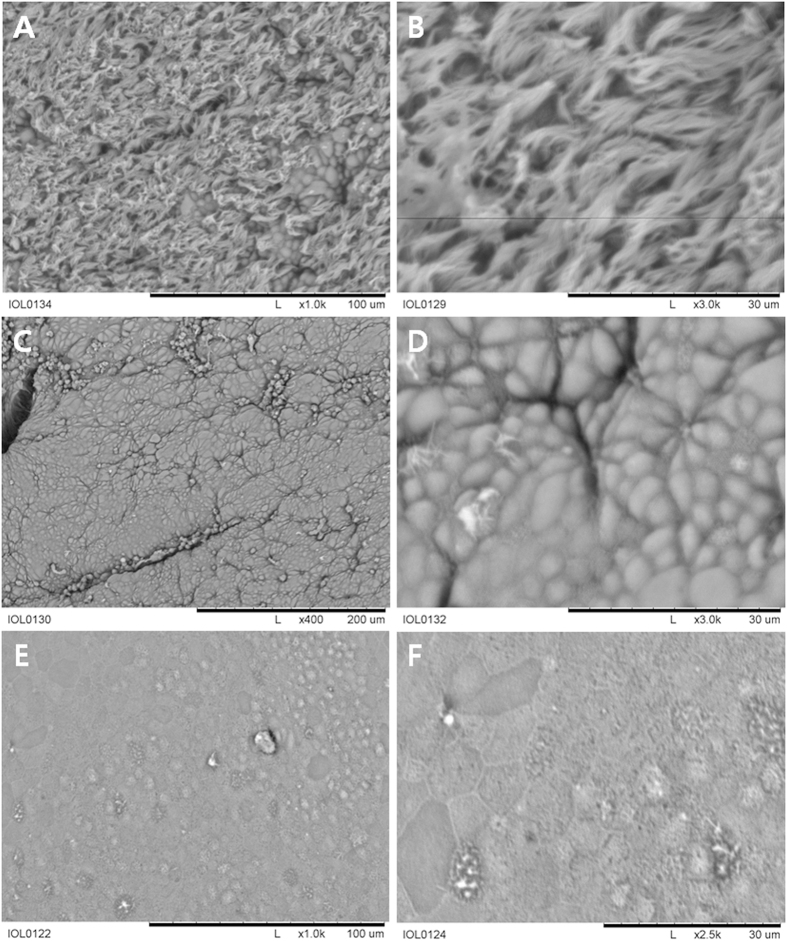
Scanning electron microscopy showing the epithelium of the trachea. (A) The surface of the normal trachea was crowded with many cilia. (B) A high-magnification image of the surface of the normal trachea revealed relatively healthy ciliated epithelium. (C) The surface of the neo-trachea near the transition zone between the normal trachea and the neo-trachea showed scanty cilia. (D) A high-magnification image of the neo-trachea surface revealed a relatively firm epithelial ultrastructure; however, ciliary structure was notably sparse. (E) In a small area of the neo-trachea, a dysmorphic surface was observed. (F) A magnified view of the dysmorphic area showed no ciliary structure.

