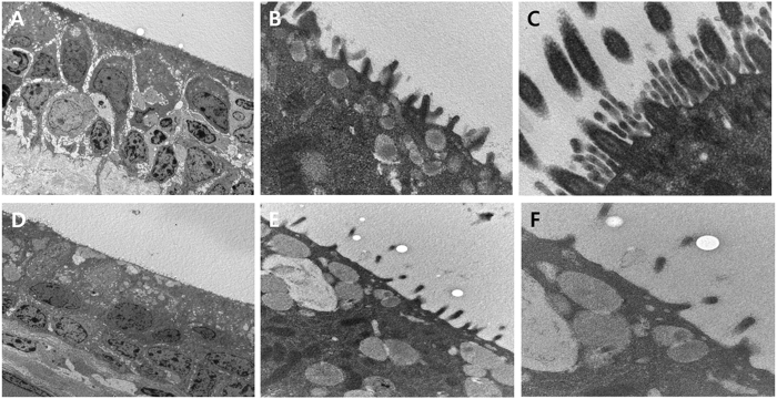
Figure 7.
Transmission electron microscopy showing the epithelium of the trachea. (A) The normal trachea revealed pseudostratified columnar ciliated epithelium. (B) Higher magnification of the normal trachea showed cilia. (C) Higher magnification of the normal trachea clearly revealed the ciliary structure. (D) The neo-trachea showed stratified squamous epithelium with scanty cilia. (E) Higher magnification of the neo-trachea indicated scanty but undoubtedly present cilia. (F) Higher magnification of the neo-trachea revealed ciliary structures.

