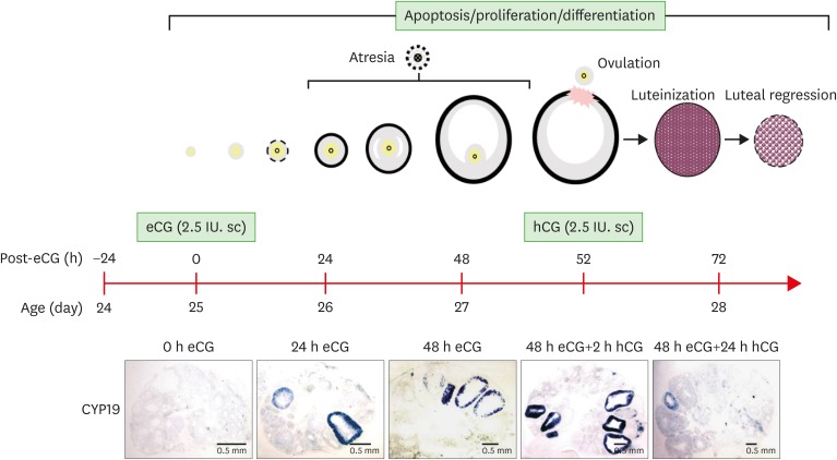
Figure 1.
Schematic of mouse ovulation model and ovarian localization of CYP19 mRNA. Ovaries were collected at 0, 24, 48 h after eCG (2.5 IU) to initiate follicular development, and 2, 24 h after hCG (2.5 IU) to induce ovulation and luteinization. The mouse ovulation model is characterized by the expected time- and cell-specific expression of CYP19 mRNA using in situ hybridization with digoxigenin CYP19 RNA probe.

