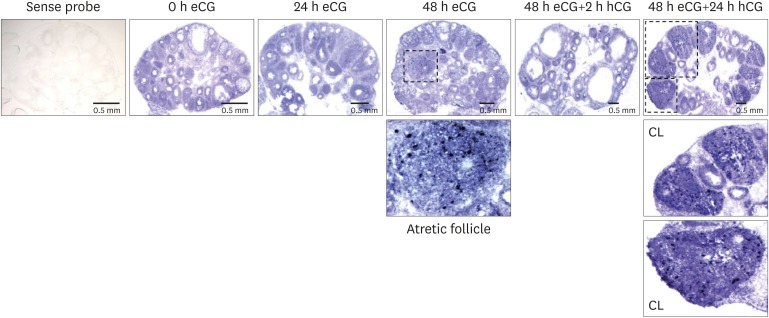
Figure 2.
Localization of SAA3 mRNA in the mouse ovary during gonadotropin-induced follicle development and ovulation. Hybridization of antisense SAA3-labeled riboprobes was visualized as purple-blue precipitate in the whole ovary at respective time points (0, 24, 48 h post-eCG injection, and 2, 24 h post-hCG injection). Incubation of tissue sections with labeled sense probe revealed no hybridization.

