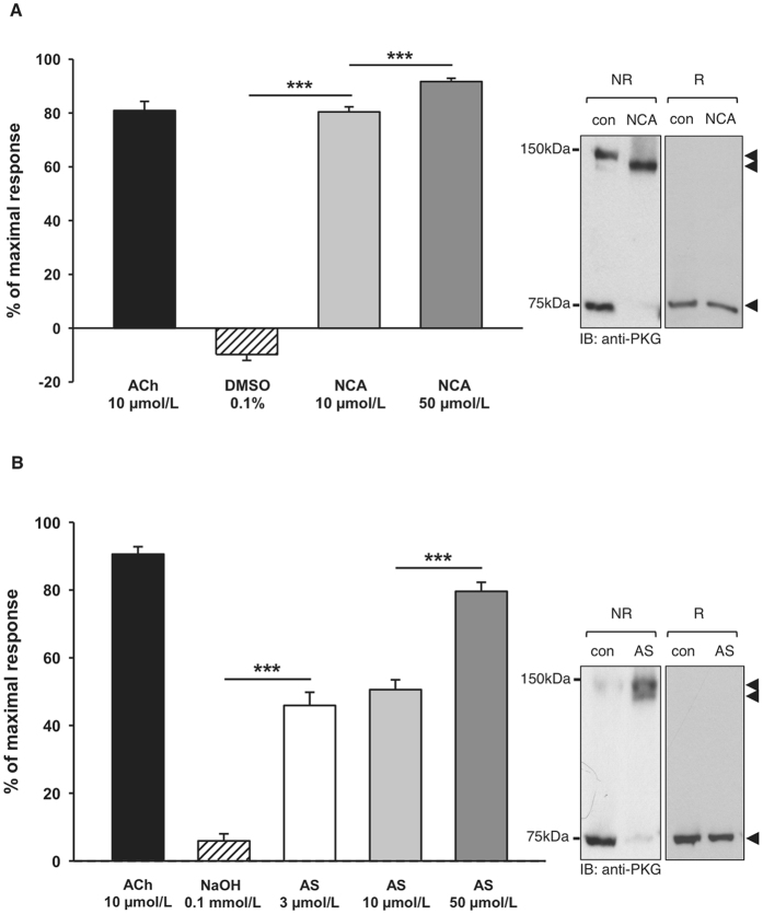
Figure 5.
Correlation of intradisulfide formation in endogenous PKGIα with HNO-mediated vasorelaxation in vivo. Both HNO donors induced concentration-dependent dilations in arterioles in vivo. (A) The effect of NCA was studied in 68 vessels from 6 mice. (B) The effect of AS was studied in 73 vessels from 6 mice. Data are given as mean ± SEM. *** indicates P < 0.001 for paired comparisons (t-test). Western immunoblot analysis for PKGIα was performed under reducing (R) and non-reducing (NR) conditions in isolated cremaster muscles exposed to 50 µmol/L NCA or AS. Black arrows indicate the positions of monomeric (75 kDa), inter- (150 kDa) and intradisulfide (below 150 kDa) PKGIα.

