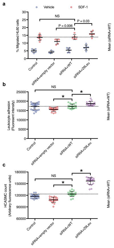
Figure 4. Functional assessment of ARHGEF26 p.Val29Leu in vitro.
a) ARHGEF26-29Leu increases leukocyte transendothelial migration. HAEC were transfected with non-targeting siRNA and empty vector (control), siRNA against ARHGEF26 3′-UTR and empty vector, siRNA and ARHGEF26-WT, or siRNA and ARHGEF26-29Leu. Transfected HAEC were plated on transwell inserts and treated with 10 ng/mL TNF-α. Differentiated HL60 cells were loaded on the upper chambers of transwells and allowed to transmigrate across HAEC towards vehicle (blue) or 50 ng/mL SDF-1 (red). The migrated cells were quantified as percentage of input cells per well (n=5 or 6; mean±s.d.; F=11.89, DF=3 by two-way ANOVA within vehicle and SDF-1 subgroups with Fisher’s LSD test; variance among vehicle subgroups non-significant; NS, not significant; representative of 3 independent experiments).
b) ARHGEF26-29Leu increases leukocyte adhesion on endothelial cells. HAEC were transfected as 2a) and cultured on 96-well plates until confluent and treated with 10 ng/mL TNF-α. Calcein-AM-labeled THP-1 cells were incubated with HAEC and washed to remove non-adherent cells. The adherent cells were lysed, quantified by Calcein-AM fluorescence and compared to siRNA+WT (n=25, 17, 20, and 17; mean±s.d.; F=14.53, DF=3 by one-way ANOVA; NS, not significant; * P<0.0001 compared to siRNA+WT; representative of 3 independent experiments).
c) ARHGEF26-29Leu increases vascular smooth muscle cell proliferation. HCASMC were transfected as 2a) and made quiescent by serum starvation for 48 h, followed by 72-h proliferation in normal serum medium. Cell proliferation was quantified by a luminescent assay and compared to siRNA+WT (n=20; mean±s.d.; F=197.5, DF=3 by one-way ANOVA; NS, not significant; * P<0.0001 compared to siRNA+WT; representative of 3 independent experiments).

