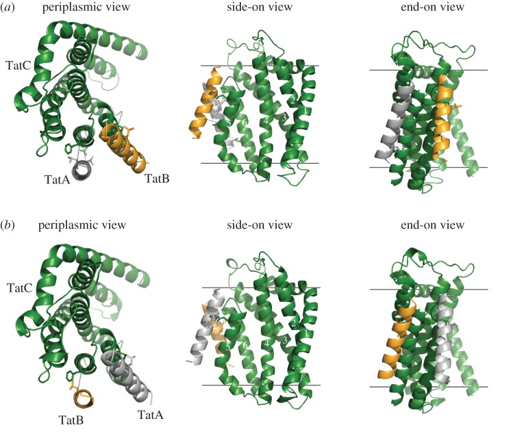
Figure 4.
Models of the TatABC trimer in the resting and activated state. Three views of (a) the resting-state TatABC complex and (b) the substrate-activated TatABC complex. TatA is shown in silver, TatB gold and TatC green. Note that in (b) the substrate signal peptide is not shown as it is currently unclear precisely where it binds in the activated state.

