
Figure 1. Muscarinic receptor stimulation causes hyperpolarisation of the RMP that is sufficient to abolish AP generation in dLGN INs.
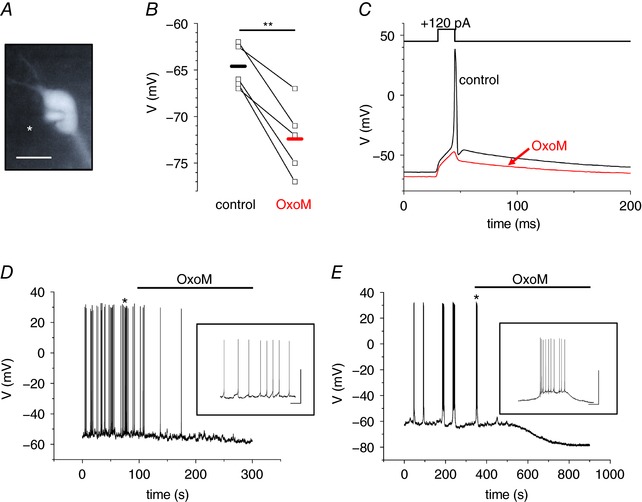
A, fluorescence image of a dLGN IN obtained from a brain slice of an EGFP‐GAD67 mouse. Note the patch pipette on the left side of the cell (marked with asterisk in image). The scale bar represents 10 μm. B, scatter diagram of the RMP under control conditions and during the application of OxoM. Points connected by lines indicate individual cell changes. The horizontal bars indicate the mean RMP under the two recording conditions (black, control; red, OxoM). C, current‐clamp recording of an IN in the absence (black trace) and presence (red trace) of OxoM (10 μm). OxoM mediates membrane hyperpolarization, which is sufficient to abolish the induction of an AP during a short lasting (15 ms) depolarizing current step (+120 pA). D and E, whole cell voltage recordings of spontaneously active INs revealing tonic firing (D) and rhythmic bursting (E). Muscarinic receptor stimulation by OxoM (10 μm; as indicated by the horizontal line) leads to membrane hyperpolarization and silencing of INs. Insets show the activity patterns at higher temporal resolution. Scale bars represent 50 mV and 2 s, respectively.