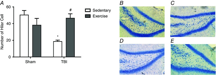
Figure 13. Previous physical training protects hippocampal cell loss after FPI.

Histological analysis (H&E staining) demonstrated previous exercise training protected against cell loss in the dentate gyrus hilus induced by FPI (A). Histological analysis in the displays: sham/sedentary group (B), sham/trained (C), TBI/sedentary (D) and TBI/trained (E) animals. Data are expressed as the mean ± SEM (n = 6–7). * P < 0.05 compared to all other groups. # P < 0.05 compared to the sedentary/TBI group. [Color figure can be viewed at wileyonlinelibrary.com]
