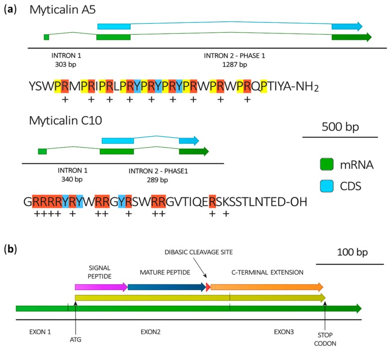
Figure 3.
(a) Gene structure of myticalin A5 and myticalin C10, deduced by the alignment of de novo assembled transcripts with the draft reference genome of Mytilus galloprovincialis [37]. The mature peptides generated by the post-translational processing of the prepropeptides encoded by the two genes are also indicated, highlighting proline (yellow), arginine (red) and tyrosine (blue) residues and indicating positive net charges at pH = 7; (b) Typical organization of myticalin mRNAs.

