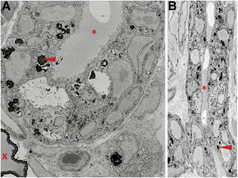
FIGURE 6.
Serial block face scanning electron microscopy (SBEM) of mammary duct from malignant carcinoma in aged retired breeder rat. Transverse (A) and longitudinal (B) views of SBEM image stack demonstrating large microcalcification (red X) and smaller, osmiophilic structures (red arrowheads) within or between mammary luminal and myoepithelial cells but not within ductal lumen (red asterisk). Images were taken at 18,500× magnification.

