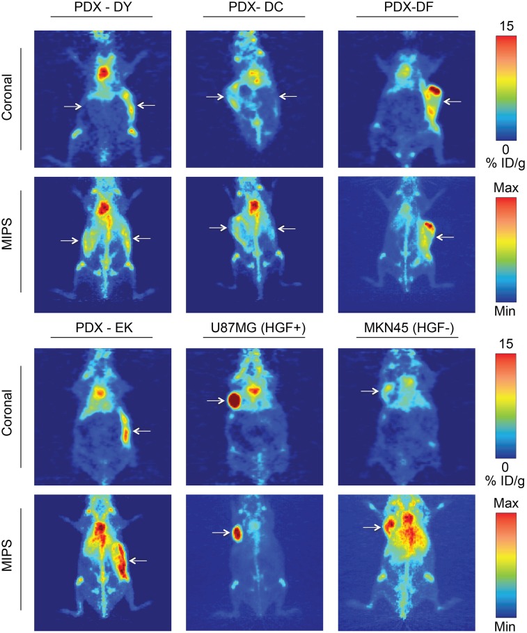
FIGURE 5.
89Zr-DFO-AMG102 (∼30 μg, ∼4.8–5.6 MBq [∼130–150 μCi], 200 μL of sterile saline) PET images 120 h after injection comparing uptake in different tumor types, showing high uptake in HGF+ U87MG tumors (∼40 %ID/g) and low uptake in HGF− MKN45 tumors (∼5–10 %ID/g). Mice bearing gastric PDXs (DY, DC, DF, EK) with previously unknown levels of HGF show similarly low uptake to the HGF− MKN45 xenografts, noninvasively determining little or no HGF present. Tumors are highlighted with white arrows, and images with 2 arrows indicate bilateral xenografts (DY, DC); full serial PET images are in Supplemental Figures 8–9.

