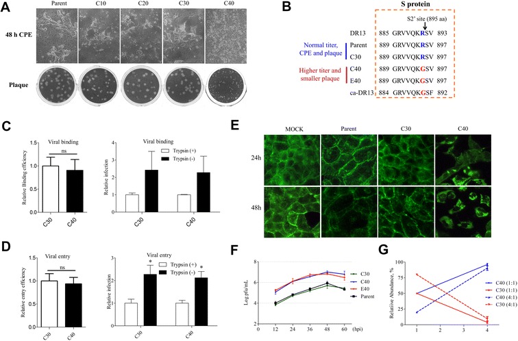
Figure 4.

The effect of mutation R895G of S protein on PEDV replication and viral fitness. (A) Comparison the CPE (48 hpi) and plaque phenotype of 85-7 parent strain (P5) with the precursor strains of C40 strain (strains C10, C20, and C30). (B) Alignment of the amino acid sequences of the N-terminal fusion peptide (FP) of DR13, ca-DR13, 85-7 parent, C30, C40, and E40 strains. The ca-DR13, and its parental strain DR13, were reference sequences. The arrow indicated the putative S2′ cleavage site. (C) The relative binding efficiency and its trypsin-independence of C30 and C40 strains. (D) The relative entry efficiency and its trypsin-independence of C30 and C40 strains. (E) Comparison of cytoskeletal reorganization in mock, 85-7 parent, C30, and C40-infected Vero cells. The infected cells were fixed and strained with FITC-Phalloidin at 24 and 48 hpi, respectively, and then photographed under a fluorescent microscope (magnification 400×). (F) Comparison of the virus curve of C30 with 85-7 parent, C40, and E40 strains. (G) Effect of the PEDV R895G mutation on viral fitness. Competition assays between the C30 mutant virus (red lines) and the C40 virus (blue lines) were performed. Vero cells were co-infected with C30 and C40 viruses at a ratio 1:1 (full line) or 4:1 (dotted line) and supernatants were passaged serially 3 times every 36 h. Error bars represent the standard deviation from three independent experiments.
