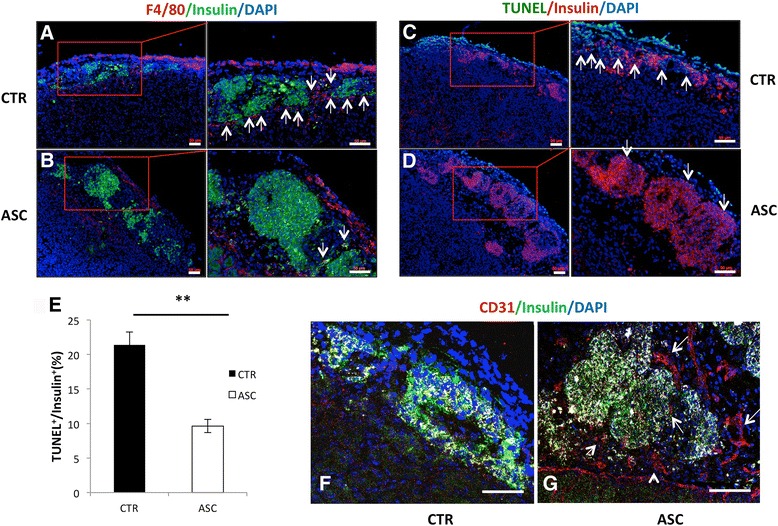
Fig. 3.

Immunohistochemical analysis of mouse islet grafts. a, b Analysis 3 days post transplant shows more macrophages and less insulin in control islets (CTR, a) compared to islets cotransplanted with CP-ASCs (ASC, b). Red, F4/80+ cells; green, insulin. Arrows point to macrophages. c, d More cell death observed in control (c) compared to CP-ASC islets (d) identified by TUNEL assay. Green, apoptotic cells; red, β cells. Arrows point to TUNEL+insulin+ cells. e Quantification of TUNEL+ among insulin + cells in control or CP-ASC cotransplanted islets. **p < 0.05, Student’s t test. f, g Tissues 10 days post transplant. Immunohistochemical staining of endothelial cells (CD31+) is less in control islets (f) compared to ASC islet grafts (g) using the anti-CD31 antibody. Red, CD31+ cells; green, insulin+ cells. Arrows point to CD31+ cells. Tissue sections from at least three individual mice for each condition were analyzed. a–e Observed using the ZEISS AxioImager M2 Imaging System. f, g Observed using a Leica SP5 confocal microscope. Scale bar = 50 μm. ASC adipose-derived mesenchymal stem cell, TUNEL terminal deoxynucleotidyl transferase-mediated dUTP nick end-labeling (Color figure online)
