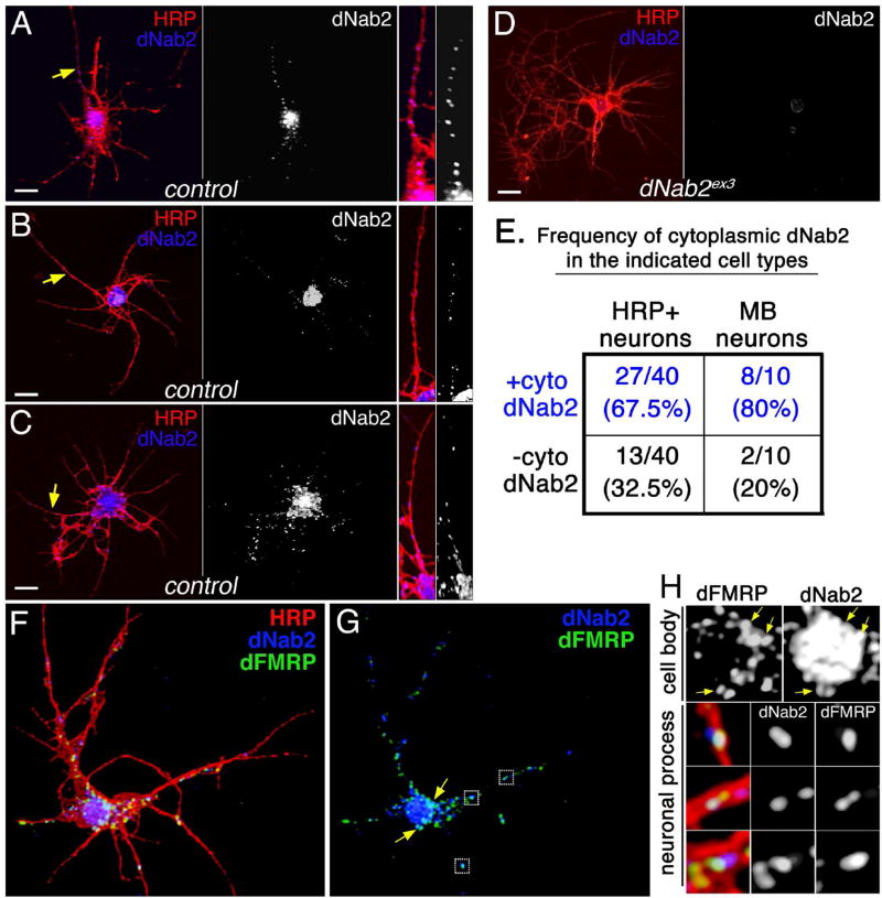
Figure 2. dNab2 colocalizes with dFMRP in mRNP-like puncta.
(A-C) Control (wt) or (D) dNab2 null (ex3) 24hr APF (after puparium formation) brain neurons cultured for 72hr and labeled with anti-HRP (red; neuronal membranes) and anti-dNab2 (blue). Scale bar=10µm. Rightmost panels in A-C are magnified views of dNab2 puncta in neurites (yellow arrows). (E) Frequency of cytoplasmic dNab2 in wt neurons (“brain neurons”; left) or Kenyon cells (“MB neurons”; right) labeled by CD8:GFP expression (CD8-GFP/+;;OK107>Gal4/+). (F-H) A single wt 24h APF brain neuron triple labeled with anti-HRP (red), anti-dFMRP (green), anti-dNab2 (blue). (G) Overlapping dNab2:dFMRP signals in the cell body (arrows) or neuronal process (boxes). (H) Magnified views of regions highlighted in G showing colocalization of dNab2 and dFMRP in the soma (“cell body”; see arrows) and processes (“neuronal process”).

