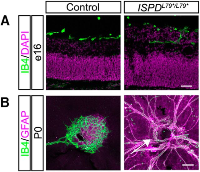
Figure 3.
Dystroglycan regulates normal retinal vasculature development. A, IB4-labeled hyaloid vasculature is present in the vitreous adjacent to the GCL in control retinas (left), but is embedded within ectopic cell clusters ISPDL79*/L79* (right) retinas at e16. B, Flat-mount retinas at P0 show the emergence of the primary vascular plexus (IB4, green) and astrocytes (GFAP, purple) in controls (left). In ISPDL79*/L79* retinas (right), the emergence of astrocytes and the primary vascular plexus into the retina is delayed (arrow) and there is a persistence of hyaloid vasculature. Scale bars: A, 50 μm; B, 100 μm.

