Figure 5.

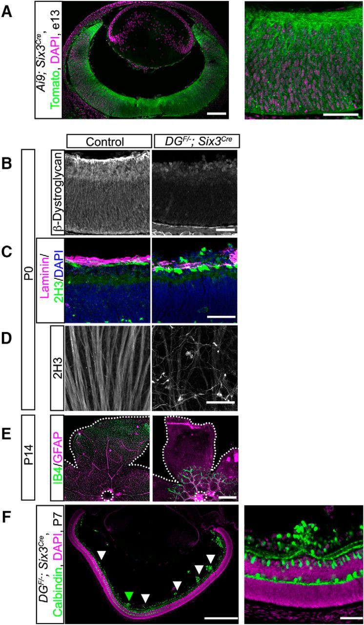
Conditional deletion of dystroglycan in the developing retina results in migration and axon guidance defects. A, Recombination pattern of Rosa26-lox-stop-lox-TdTomato;Ai9 reporter (green) by Six3Cre shows expression throughout the retina and in axons at e13. B, Dystroglycan protein expression is lost in DGF/−;Six3Cre mice. C, D, DGF/−;Six3Cre (right) mice exhibit ILM degeneration (top, purple, laminin) and abnormal axonal fasciculation and guidance (top, green, bottom, 2H3). E, Primary vascular plexus (IB4, green) and astrocytes (GFAP, purple) migrate from the optic nerve head (dashed circle) to the edge of the retina (dashed line) in control retinas at P14 (right). Vascular and astrocyte migration is stunted in DGF/−;Six3Cre retinas. F, Focal migration defects (arrowheads) in P7 DGF/−;Six3Cre retinas are present across the entire span of the retina. Green arrowhead indicates high-magnification image in F (right). Scale bars: A, left panel, 100 μm, right panel 50 μm; B–D, 50 μm; E, 500 μm; F, left panel 500 μm, right panel 50 μm.
