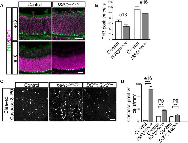
Figure 9.
Loss of dystroglycan results in increased developmental cell death. A, Immunohistochemistry for mitotic cells (PH3) at e13 (top) and e16 (bottom) shows normally positioned mitotic retinal progenitor cells adjacent to the RPE. B, Quantification of mitotic cells shows no difference between control and ISPDL79*/L79* mutants (p > 0.05, t test, n = 4 control and 4 mutant retinas at e13, p > 0.05, t test, n = 4 control and 4 mutant retinas at e16). C, D, Immunohistochemistry for cleaved caspase-3 in a flat-mount preparation of P0 DGF/−;Six3Cre retinas shows an increase in apoptotic cells (p = 0.0279, t test, n = 18 samples from 6 control retinas, 17 samples from 6 mutant retinas). D, Quantification of cleaved caspase-3-positive cells shows an increase in apoptotic cells at e16 (p < 0.0001, t test, n = 18 samples from 6 control retinas, 18 samples from 8 mutant retinas) and P0 (C, D; p < 0.0001, t test, n = 18 samples from 6 control retinas, 15 samples from 5 mutant retinas) between control (left) and ISPDL79*/L79* (middle) retinas. Scale bar, 50 μm.

