. 2017 Aug 15;6:e27960. doi: 10.7554/eLife.27960

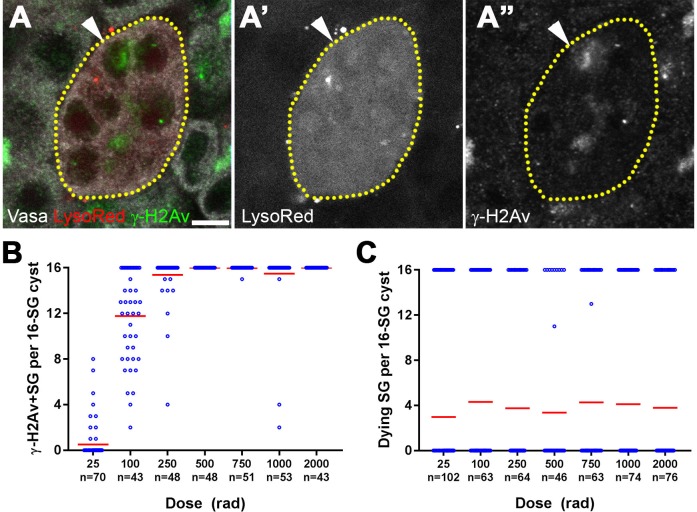
Figure 2. All SGs within a cyst die even when only a fraction of cells exhibit detectable DNA damage.

(A) An example of a dying 16-SG cyst (yellow dotted outline) with only a subset of SGs containing detectable DNA damage (arrowhead). γ-H2Av (green), Lysotracker (red), Vasa (white). Bar: 5 µm. (B) Number of γ-H2Av-positive cells within each 16-SG cyst at various radiation doses. Blue circles, individual data points. Red line, mean. n = number of 16-SG cysts scored. (C) Number of Lysotracker-positive cells within each 16-SG cyst. Blue circles, individual data points. Red line, mean. n = number of 16-SG cysts scored.

Figure 2.

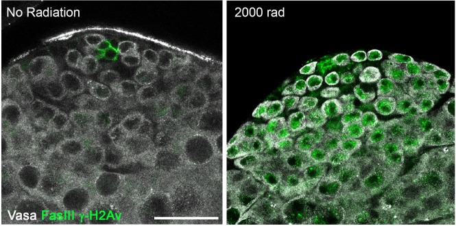
Figure 2—figure supplement 1. γ-H2Av can be strongly detected in germ cells following irradiation.

Figure 2—figure supplement 1.

Representative images of testes apical tips from unirradiated and irradiated flies. Vasa (white), FasIII and γ-H2Av (green). Bar: 25 µm.
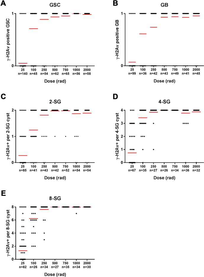
Figure 2—figure supplement 2. All SG stages show gradual accumulation of γ-H2Av-positive cells with increasing radiation.

Figure 2—figure supplement 2.

Number of γ-H2Av-positive cells per cyst at increasing radiation doses in (A) germline stem cells (B) gonialblasts (C) 2-SG (D) 4-SG (E) 8-SG. Red line, mean. n = number of 16-SG cysts scored.