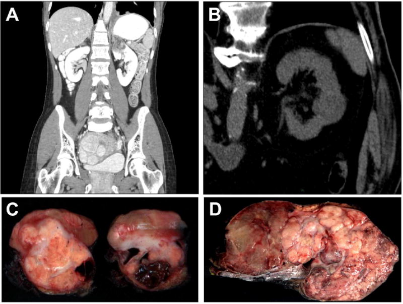
Figure 1. Clinical and Gross Features of TC-PD.

A. Contrast abdominal CT scan in the coronal plane of case TC-PD 17 demonstrates a 4 cm solid and cystic tumor in the upper pole of the left kidney. The lower left lung demonstrates pleural-based nodularity and effusion, which proved to be metastatic disease, while uterine leiomyomatosis was quite prominent in this case, which was FH-deficient immunohistochemically. B. Non-contrast abdominal CT scan of case TC-PD 7 demonstrates a lesion in the posterior of interpolar left kidney which proved to be a TC-PD with FH-retained immunophenotype. C. Gross photograph of representative cross sections of the nephrectomy specimen from case TC-PD 17, which shows a solid (predominant on left section) and cystic (predominant on right section) tumor appearing based in the medulla. D. Gross cut section of case TC-PD 3, where tumor is seen diffusely infiltrative of the entire kidney. The areas to the left show some features of the multicystic so-called “bubble wrap” appearance described for conventional tubulocystic carcinoma.
