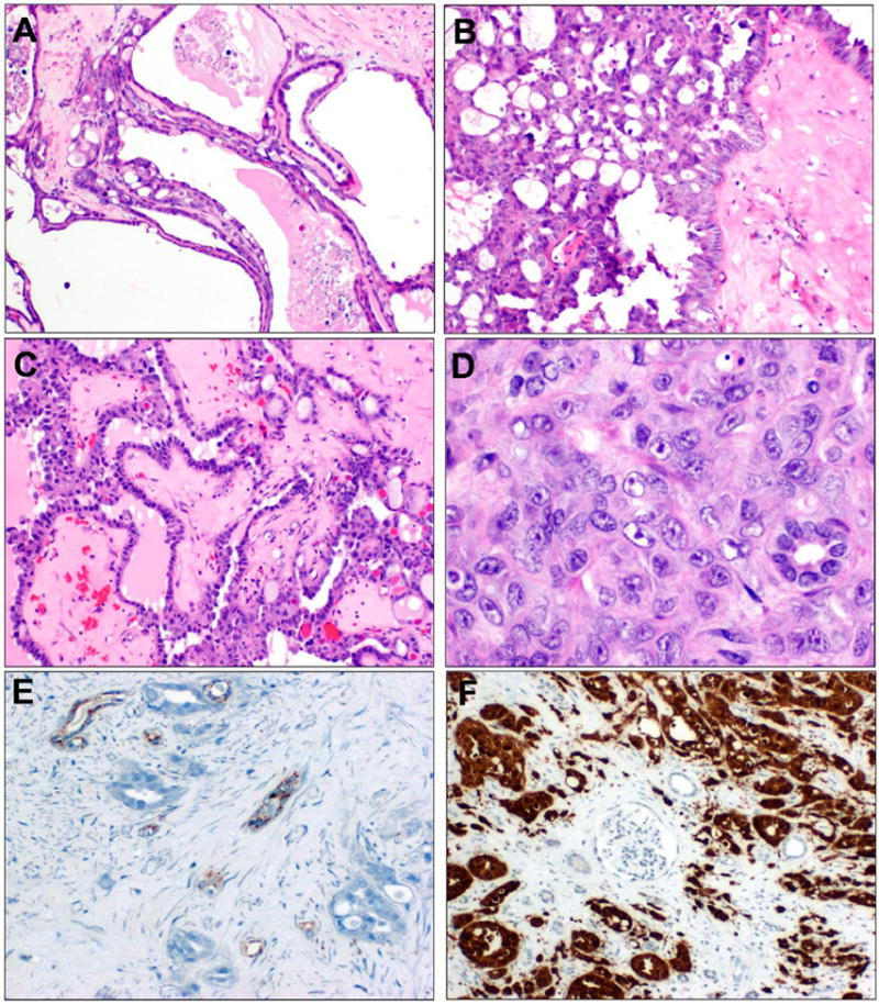
Figure 3. TC-PD with FH-deficient immunophenotype.

Case TC-PD 7 also demonstrated more focal TC-PD morphology, with these areas seeming to give rise to infiltrative glands with desmoplasia (A). More prevalent growth patterns included cribriform growth (B), foci of intracystic papillary growth with hyalinized cores (C), and solid areas, with prominent nucleoli and examples of focal perinucleolar clearing (D). By immunohistochemistry, this carcinoma was negative for FH (panel E, note internal positive control entrapped tubules) and diffusely positive for 2SC in nucleocytoplasmic manner (panel F, note entrapped internal negative control glomerulus and tubules).
