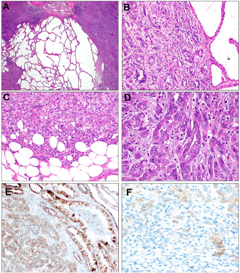
Figure 4. TC-PD with FH-retained immunophenotype.

Case TC-PD 27 showed areas of tubulocystic growth directly juxtaposed to dense, cellular areas of poorly differentiated morphology (A). On higher power, the infiltrative glandular morphology can be seen directly juxtaposed to tubulocystic areas (B). At the periphery, solid and nested patterns are seen invading into perinephric adipose (C). Most of the tumor showed infiltrative, collecting duct carcinoma-like glands seen embedded in inflamed desmoplastic stroma (D). This case showed diffuse expression of FH by IHC (E). 2SC showed only focal, weak cytoplasmic staining (1+, F), which is interpreted as negative for this stain.
