
Figure 5. Inhibition of BLA→InsCtx neurons attenuates food cue responses in InsCtx.
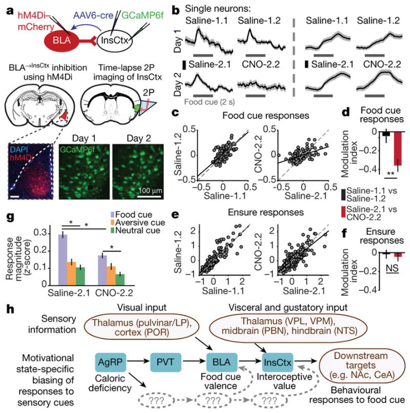
a, Inhibition of BLA→InsCtx neurons while imaging InsCtx. Brain schematics were adapted with permission from ref. 43. b, Example single neurons. Scale bars, 0.2 normalized ΔF/F. c, Average cue-evoked responses for food-cue-responsive neurons. Dots, individual neurons. Linear regression line, black; line of unity, dashed grey. d, Modulation index. Median ± s.e. median. **P = 0.001, Mann–Whitney U-test, n = 98 neurons, four mice. e, Average Ensure-evoked responses. f, Modulation index. Median ± s.e. median. NS, P = 0.5; Mann–Whitney U-test, n = 176 neurons, four mice. g, Cue response magnitude. Within condition, *P < 3.9 × 10⁻⁷, Kruskal–Wallis test. Saline-2.1: food versus aversive, P = 6.2 × 10⁻⁹; food versus neutral, P = 4.2 × 10⁻⁹; aversive versus neutral, P = 0.6; CNO-2.2: food versus aversive, P = 1.5 × 10⁻⁴; food versus neutral, P = 5 × 10⁻⁹; aversive versus neutral, P = 0.2; Saline-2.1 versus CNO-2.2: food, P = 1.7 × 10⁻⁸; aversive, P = 0.04; neutral, P = 0.01, Mann–Whitney U-test (n = 118, 57, and 36 food, aversive, and neutral cue-responsive neurons, respectively, four mice). h, Pathway from AgRP neurons to InsCtx. Grey, additional unknown pathways (Supplementary Discussion). LP, lateral-posterior thalamus; POR, postrhinal cortex; VPL, ventroposterior lateral thalamus; VPM, ventroposterior medial thalamus; PBN, parabrachial nucleus; NTS, nucleus of the solitary tract.