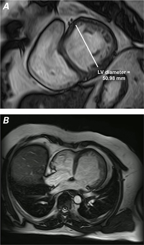
Fig. 3.

Cardiac magnetic resonance images. A) After 2 years, the short-axis, white-blood view shows decreased dilation and hypokinesia of the left ventricle (LV) at end-diastole. B) The 4-chamber, white-blood view shows improved LV contractility.
Supplemental motion images are available for Figure 3A and Figure 3B.
