Figure 5.
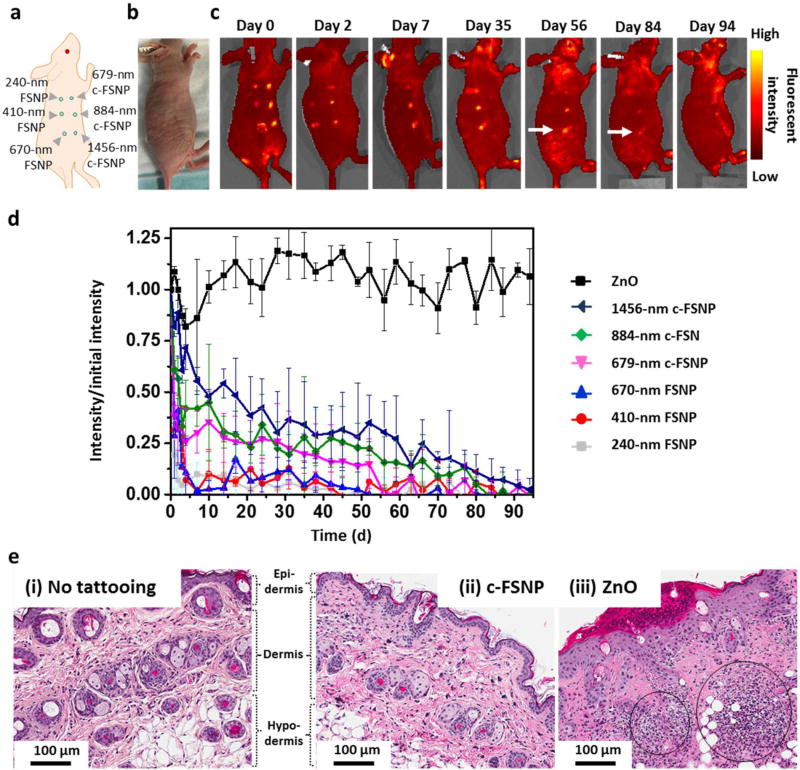
Size-dependent intradermal retention study of FSNPs and c-FSNPs. (a) Three different-sized c-FSNPs (i.e., 679, 884, and 1456 nm) along with three different-sized FSNPs (i.e., 240, 410, and 670 nm), each containing 0.75 µg of MPS-PPV, are tattooed at six different locations on the back of the nu/nu mice (n = 3). (b) Photograph of a mouse tattooed with various-sized FSNPs and c-FSNPs under ambient light irradiation. (c) Fluorescent images of the tattooed mouse obtained using the in vivo optical imaging system (excitation/emission = 465/ 520 nm; exposure time = 2 s), for 94 days. (d) Time-dependent fluorescent signal of tattoo pigments, including a commercially available pigment (i.e., ZnO), FSNPs, and c-FSNPs. The fluorescent signals are normalized to the initial fluorescent signal intensity at day 0. In a long-term fluorescent signal observation up to 94 days, both FSNPs and c-FSNPs display a size-dependent fluorescent signal with a finite intradermal retention time. Among various FSNPs and c-FSNPs, 1456 nm sized c-FSNPs (arrow in panel c) show a fluorescent signal up to 84 days with the slowest fluorescent signal decay. The commercially available tattoo pigment, ZnO, exhibits a consistent fluorescent intensity over 94 days. (e) H&E stained skin sections from a nu/nu mouse tattooed with 1456 nm sized c-FSNPs after 2 days post-tattooing (100× magnification). Compared to (i) normal skin without tattooing, (ii) no obvious inflammation cells were observed in the skin of nu/nu mouse after c-FSNP tattooing. (iii) In contrast, commercially available tattoo pigments (i.e., ZnO) attract a large population of inflammation cells (black circles).

