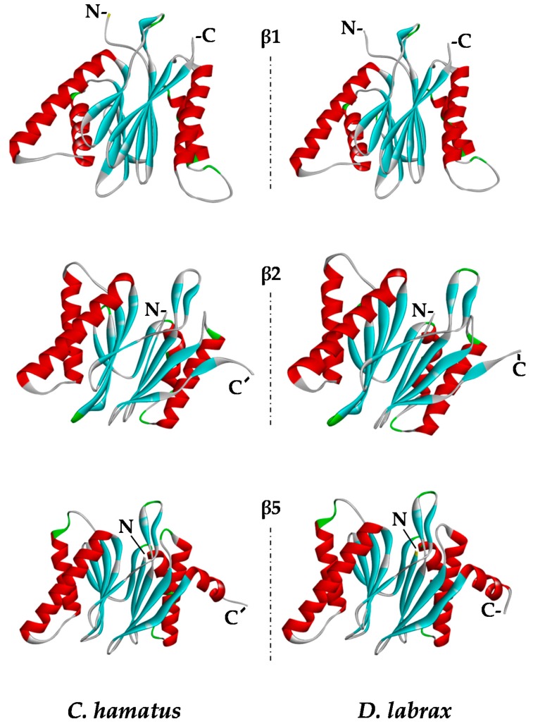
Figure 7.
Structural models of β1, β2, and β5 proteasome subunits. α Helix, β-strand and turn structures are indicated with red, cyan and green colors, respectively. N- and C-termini are indicated by N- and C-, respectively. Models were generated as described in the Materials and Methods section. Images were created with Discovery Studio software.

