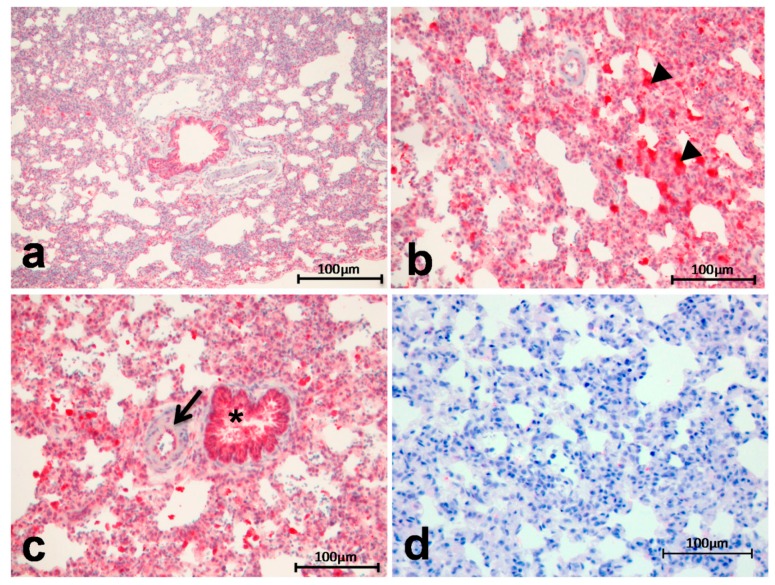
Figure 2.
Arg I showed an increased expression and tissue deposition in induced pulmonary hypertension (IPH) rats (b–c) compared to the controls (a) enhanced staining positivity in the lung parenchyma and in alveolar macrophages ((b), arrowheads = alveolar macrophages) and mild to moderate positivity in endothelial cells of peribronchial arteries ((c), arrow); staining intensity of the epithelium of the bronchioles is more or less equal to that occurring in normal tissue ((c), *); negative control for Arg I staining (d).

