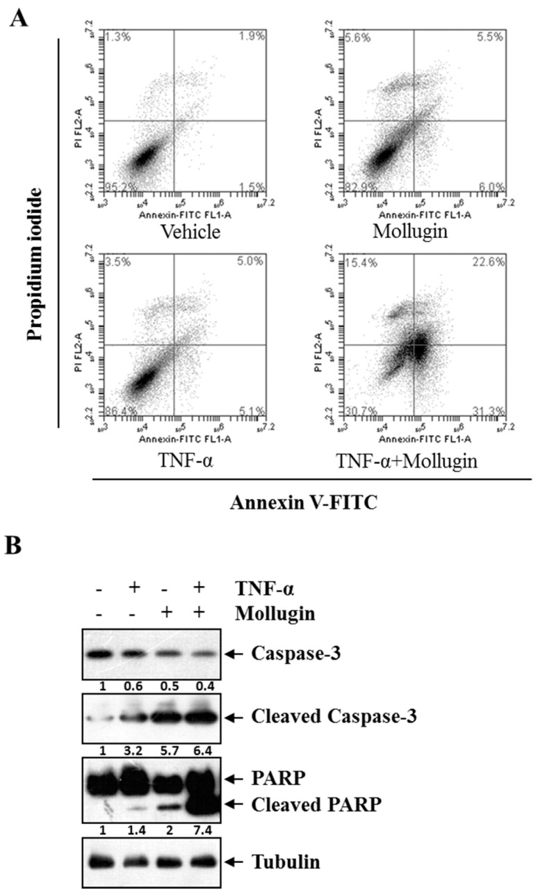
Figure 4.
Mollugin potentiates TNF-α-induced apoptosis. (A) HeLa cells were pre-incubated in the presence of 80 μM mollugin for 12 h prior to treatment with 10 ng/mL TNF-α for 24 h. Then, the cells were collected and labelled with Annexin V-FITC and propidium iodide for staining. The stained cells were analyzed using a flow cytometer. The lower right quadrant represented the early apoptotic cell (Annexin-V+ and PI−) and the upper right quadrant represented the late apoptotic cells (Annexin-V+ and PI+); (B) HeLa cells were pre-incubated in the presence of 80 μM mollugin for 12 h prior to treatment with 10 ng/mL TNF-α for 24 h. Western blot assay was performed for the total and cleaved capase-3, PARP. Tubulin was used as an internal control.

