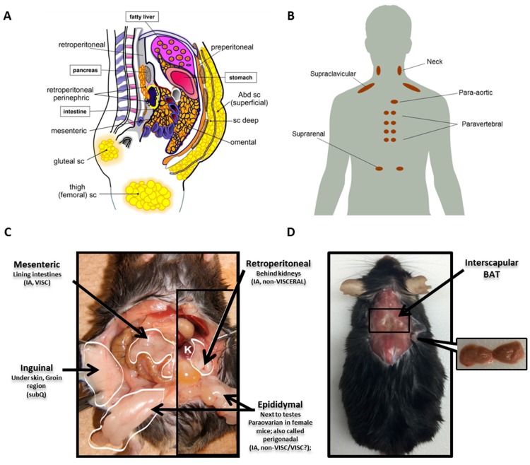
Figure 1.
Distribution of white AT (WAT) and brown AT (BAT) in humans and mice. (A) Prominent human WAT depots are shown. Subcutaneous (subQ) depots most often studied in humans include two distinct upper body depots on the ventral side, superficial and deep, as well as gluteal and femoral fat pads. Several depots are defined as intraabdominal, including perinephric around the kidneys, retroperitoneal in the retroperitoneal space behind the kidneys and mesenteric and omental WAT lining organs of the digestive tract. (B) Several human BAT depots are depicted. BAT in adult humans is present in several different locations, including the neck, heart, spinal cord and kidneys. However, the majority is contained within the supraclavicular depot. (C) Four commonly-studied WAT depots in a male mouse are shown. Inguinal fat is a subQ depot that lies just beneath the skin and is similar to the gluteofemoral subQ in humans. Intraabdominal depots include mesenteric fat associated with the intestine, retroperitoneal behind the kidneys (K) and epididymal WAT next to the testes (paraovarian surrounds the ovaries of female mice; collectively, epididymal and paraovarian are also called perigonadal). According to the stricter rules of nomenclature, only mesenteric fat is a true visceral depot in mice, as it is the only one that drains into the portal vein. (D) The location of the interscapular BAT depot in mice, the only dissectible BAT fat pad, is shown. Abd sc: Abdominal subcutaneous; IA: Intraabdominal; VISC: Visceral. Figure 1A is adapted with permission from [1]. Copyright 2013 Elsevier. Figure 1B is adapted with permission from [35]. Copyright 2010 Wiley-Blackwell. Figure 1C is adapted with permission from [36,37]. Copyright 2011 Elsevier. Figure 1D is adapted with permission from [38]. Copyright 2017 John Wiley & Son.

