Figure 2.
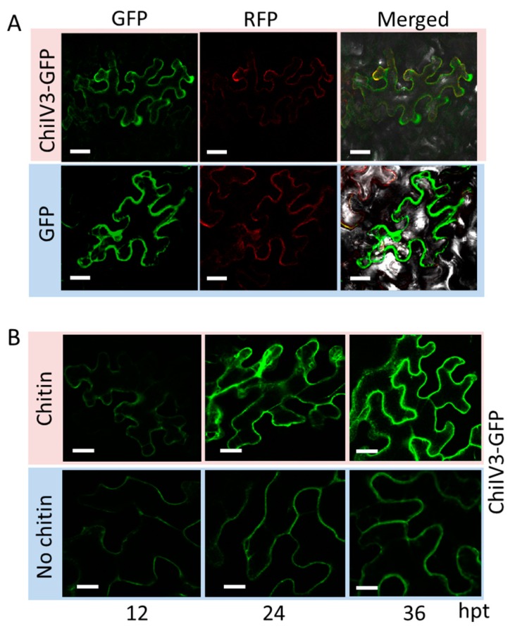
ChiIV3 was located to the plasma membrane and was activated by exogenously applied chitin. (A) Confocal images showed representative N. benthamiana leaf epidermal cells transiently expressing ChiIV3-GFP and SRC2-1-RFP both driven by 35S promoter. The fluorescent signals were detected with the confocal microscope DM6000 CS (Leica, Solms, Germany) at 48 h after Agro-infiltration. Bars = 50 μm; (B) ChiIV3 activation during MAMP (chitin) triggered immunity. Time-lapse imaging of pChiIV3:ChiIV3-GFP expressed in N. benthamiana leaves. After chitin treatment, the fluorescent signals were detected at 12 h intervals. Hpt, hours post treatment. Bars = 50 μm.

