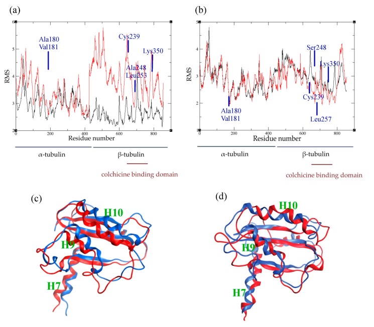
Figure 5.
Comparison between the holo and apo forms of tubulin. (a,b): average atomic fluctuations per residue in human (a) and in C. autumnale (b) tubulins. The tubulin heterodimer is represented in red in the presence of colchicine, and in black in the absence of colchicine. Key amino acids involved in colchicine binding are depicted on the graphs. (c,d): overlay of the conformations of the colchicine-binding domain of β tubulin in the presence and absence of colchicine. The conformations of the intermediate domain of the human β-tubulin are shown in (c) and the ones of the C. autumnale β tubulin are depicted in (d). The holo form of tubulin bound to colchicine is colored in blue, and the apo form in red.

