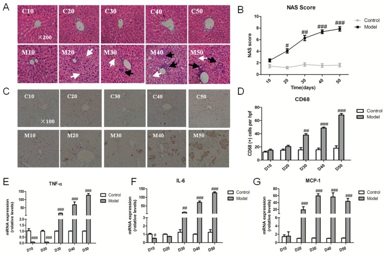
Figure 3.
High fat-sucrose diet promoted inflammation development in the liver. The hepatic inflammation change of SD rats induced by a high fat-sucrose diet on day 10, 20, 30, 40, 50. (A) Typical hematoxylin–eosin (HE) staining results of each group of rats (×200). White and black arrows display fat vacuole of hepatocytes and infiltration of inflammatory cells, respectively; (B) NAFLD activity score (NAS) of each group of SD rats after Non-alcoholic steatohepatitis (NASH) induction; (C) CD68 positive Kupffer cell (×100); (D) Semi-quantitative analysis of the CD68 positive Kupffer cell. The relative inflammation factors of the gene TNF-α (E), IL-6 (F), MCP-1 (G) mRNA expression. The values are shown as the means ± SEM (n = 6). Compared with the corresponding control group, # p < 0.05, ## p < 0.01, ### p < 0.001.

