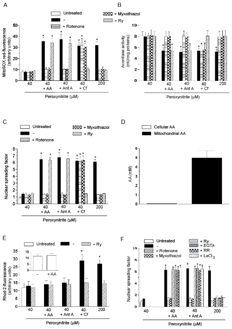
Figure 1.
The enhancing effects of AA in U937 cells exposed to low concentrations of peroxynitrite: evidence for a Ca2+-independent mechanism. (A–C) The cells were treated as shown in the figure and then exposed for: 10 min (A,B); or 30 min (C) to the indicated concentrations of peroxynitrite. AA (3 µM) was given to the cells 15 min prior to peroxynitrite. Pre-exposure to antimycin A (Ant A, 1 µM), or Cf (10 mM), was instead of only 5 min. In some experiments, rotenone (0.5 µM), myxothiazol (5 µM) or Ry (ryanodine) (20 µM) were added to the cells 5 min prior addition of AA, Ant A or Cf. After treatments, the cells were analyzed for: MitoSOX red-fluorescence (A); aconitase activity (B); and DNA strand scission (C); (D) the cells were exposed for 15 min to AA and immediately analyzed for their cellular and mitochondrial content of the vitamin; (E) Rhod 2-AM (acetoxymethyl) pre-loaded cells were treated for 10 min, as indicated in the figure, and analyzed as detailed in the Materials and Methods Section. The inset shows the Rhod 2-fluorescence response observed in cells exposed for 15 min to 0 or AA; (F) cells were permeabilized with digitonin and exposed for 10 min to the indicated concentrations of peroxynitrite alone or associated with rotenone, myxothiazol, Ry, EGTA (ethylene glycol-bis(β-aminoethylether)-N,N,N′,N′-tetraacetic acid) (10 µM), RR (ruthenium red) (200 nM) or LaCl3 (100 µM). In some experiments, the cells were exposed to AA prior to permeabilization. In other experiments, the cells were supplemented with Ant A immediately after permeabilization. Results represent the means ± SD calculated from at least three separate experiments. * p < 0.001 as compared to untreated cells (one-way ANOVA followed by Dunnett’s test).

