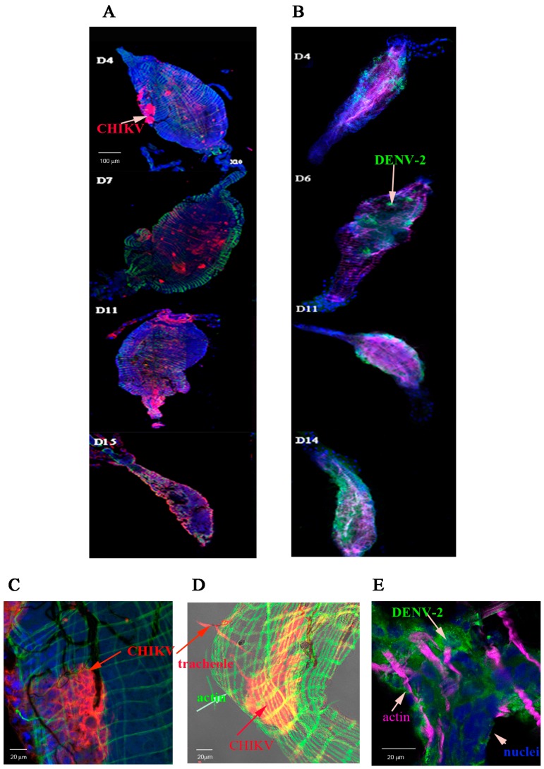
Figure 1.
Distribution of viral antigens in midguts infected by chikungunya virus (CHIKV) and dengue virus serotype 2 (DENV-2). Midguts (MG) were collected at days 4, 7, 11, and 15 post-virus exposure for CHIKV and 4, 6, 11, and day 14 post-virus exposure for DENV-2 as indicated in panels (A,B) respectively. Viral antigens were labelled using anti-CHIKV 3E4 (panel (A) in red) or anti-DENV-2 4E11 (panel (B) in green) monoclonal antibodies. Nuclei were stained with DAPI and the actin network with Alexafluor 488 (panel (A) in green) or 633 (panel (B) in magenta) phalloidin. The distribution of CHIKV antigen in a CHIKV-infected midgut at day 3 post-virus exposure was shown in panels (C,D). Muscular tissue stained in green with Alexafluor 488 phalloidin (C,D); CHIKV antigens labelled in red with Cy3-labelled 3E4 anti-E protein (C,D) and nuclei in blue (DAPI) (C). (D) shows a surperimposition of a transmission light picture to an immunofluorescence picture. The distribution of DENV-2 antigens in the posterior part of the midgut at day 3 post-virus exposure was shown in panel E. The muscular tissue is shown in magenta (Alexafluora 633 phalloidin), viral antigens in green (4E11 monoclonal antibody) and nuclei in blue (DAPI).

