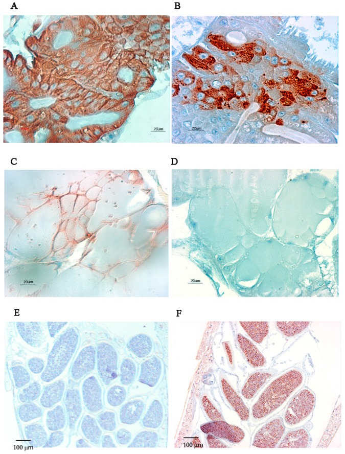
Figure 6.
Distribution of CHIKV and DENV-2 antigens in mono-infected MG, SG and eggs by IHC at 7 dpve. (A,B) MG of CHIKV and DENV-2 infected mosquitoes, respectively, Scale bar = 20 µm; (C,D) SG of CHIKV and DENV-2 infected mosquitoes, respectively, Scale bar = 20 µm; (E,F) Eggs of CHIKV and DENV-2 infected mosquitoes, Scale bar = 100 µm. Antigens are stained brown.

