Figure 1.
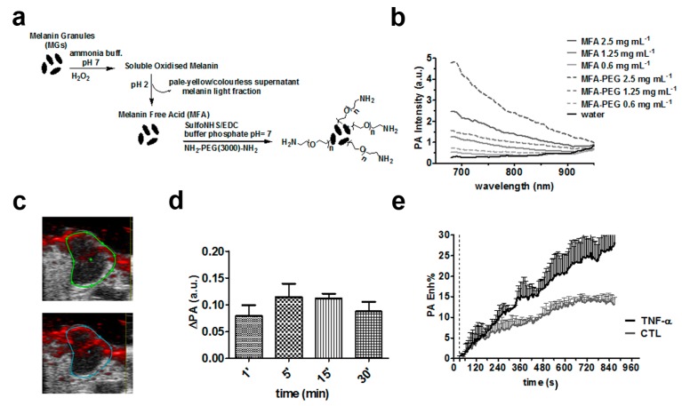
(a) Schematic illustration of the preparation of melanin-free acid derivatives (MFA) and their PEGylated form (MFA-PEG); (b) optoacoustic (OA) emission spectra of MFA and MFA-PEG at several concentrations in 1× PBS and pH = 7.4; (c) representative optoacoustic images in transverse section of tumor before (top) and 5 min after (bottom) intravenous injection of MFA-PEG (concentration: 2.5 mg/mL, volume: 0.1 mL) at 700 nm; (d) average OA signal changes in breast tumor-bearing mice (n = 4); and (e) averaged dynamic contrast-enhanced OA curves (OA Enh %) upon MFA-PEG tail vein injection for the control (n = 3) and TNF-α treated (n = 4) group, showing increased OA enhancement following antiangiogenic treatment. Reprinted with permission from reference [31], Copyright 2016, John Wiley and Sons.

