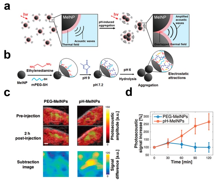
Figure 2.
(a) Schematic illustration of the proposed aggregation-induced amplification of the OA signal from melanin nanoparticles (MelNP) upon laser irradiation (hv, dashed arrow); (b) the surface modification of the bare melanin nanoparticles with citraconic amide results in the aggregation, under mildly acidic conditions (pH-MelNP); (c) combined ultrasound (US, in gray) and optoacoustic (OA, in red and yellow) images of B16 melanoma in living mice before and after the intravenous injections (concentration: 1 mg/mL, volume: 0.2 mL) of the PEG-MelNP (left column images) and the pH-responsive melanin-nanoparticles (right column images). The difference image was obtained by subtracting OA signals in the tumor site (a white box) of the pre-injection image from those in the post-injection image; (d) a change in the difference of OA signal intensity between the pre- and the post-injection images acquired at 30, 60, 90, and 120 min. Reprinted with permission from reference [32], Copyright 2016, Royal Society of Chemistry.

