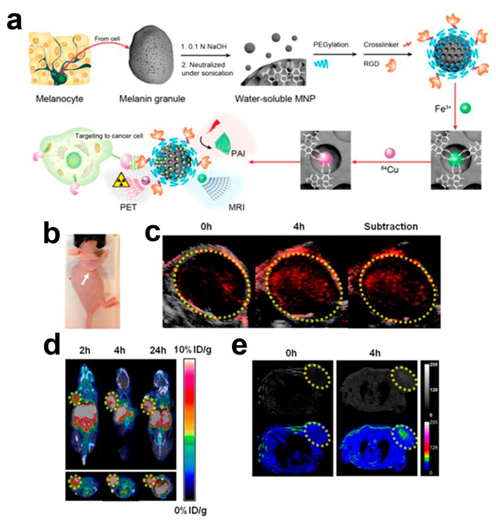
Figure 4.
(a) Schematic illustration of the synthesis of the multimodality melanin nanoparticles (MNP). After PEG surface-modification, RGD peptides were further attached to the melanin-nanoparticles for tumor targeting. Then Fe3+ and/or 64Cu2+ were chelated to the obtained MNPs for OA/MRI/PET multimodal imaging; in vivo multimodality imaging in U87MG tumor-bearing mouse: (b) photographic image (white arrow refers to the tumor position); (c) overlaying of ultrasonic (gray) and optoacoustic (red) imaging; (d) overlaying of decay-corrected coronal (top) and transaxial (bottom) small animal CT (gray) and PET (color) images; (e) MRI images (top row shows black and white images, and bottom row shows the pseudocolored images) after tail vein injection 64Cu-Fe-RGD-PEG-MNP (tumor region is enveloped by yellow dotted line). Reprinted with permission from the reference [33], Copyright 2014, American Chemical Society.

