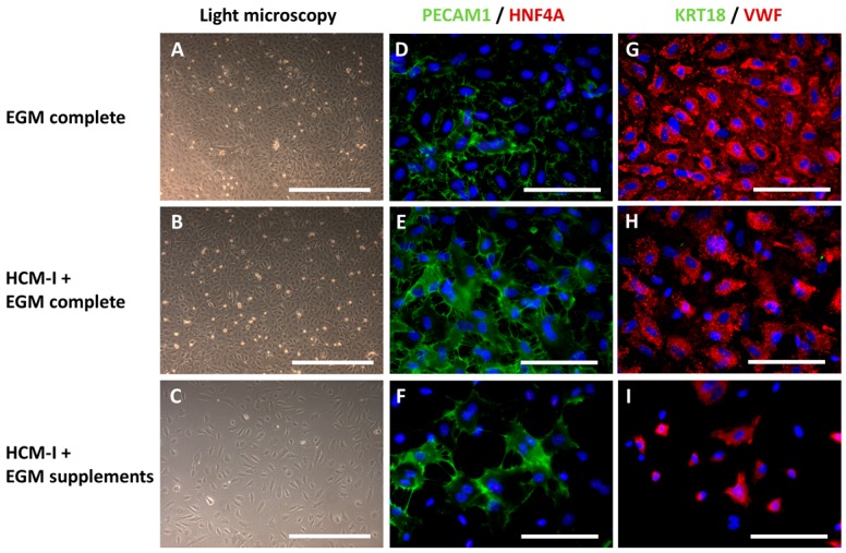
Figure 3.
Light microscopy and immunocytochemical staining of mono-cultures of human umbilical vein endothelial cells (HUVEC) after cultivation over 14 days in endothelial cell growth medium, consisting of basal medium and supplements (EGM complete), hepatocyte culture medium and EGM complete mixed at a ratio of 1:1 (HCM-I + EGM complete) or HCM enriched with endothelial cell growth supplements (HCM-I + EGM supplements). The pictures show light microscopic photographs (A–C), staining of the endothelial cell marker platelet endothelial cell adhesion molecule 1 (PECAM1) and the hepatocyte marker hepatocyte nuclear factor 4 α (HNF4A) (D–F), staining of the hepatocyte marker cytokeratin 18 (KRT18) and the endothelial cell marker von Willebrand factor (VWF) (G–I). Nuclei were counter-stained with Dapi (blue). Scale bars correspond to 500 µm for light microscopy and to 100 µm for immunofluorescence.

