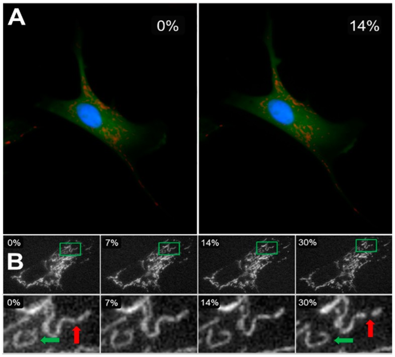
Figure 3.
Cells were cultured on elastic membranes that could be stretched equibiaxially in a stretching device. (A) A cell labeled for cytosol (green), mitochondria (red: tetramethylrhodamine methyl ester, TMRM), and nucleus (blue) at 0% (left) and 14% (right) strains. (B) The top row shows the mitochondrial network of an entire cell imaged during constant strain application at increments of 0, 7, 14, and 30% change in membrane surface area. The bottom row shows the zoomed-in details of an individual cluster (green rectangle in top row) changing shape as higher strains are applied (green arrow), as well as a cluster undergoing fission and splitting into two smaller clusters (red arrow) [77].

