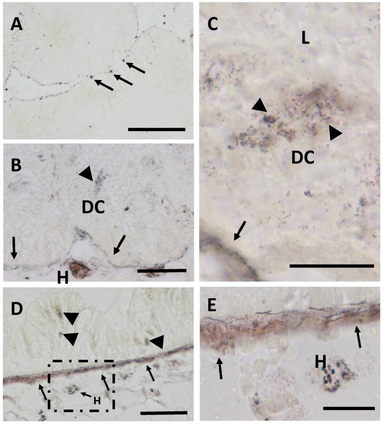
Figure 2.
Autometallography staining of the midgut gland of winkles. (A) Controls, black silver deposits (BSD) in the basal lamina (arrows) of midgut gland tubuli (scale bar: 100 µm); (B) CdH (21 days), BSD in digestive cell lysosomes (arrowheads) and in the basal lamina (arrows) of midgut gland tubules. Note the presence of BSD within hemocytes (H) of the interstitial connective tissue (scale bar: 50 µm); (C) CdH (21 days), detail of BSD in digestive cell (DC) lysosomes (arrowheads) near the tubular lumen (L) and in the basal lamina (arrows) of a midgut gland tubule (scale bar: 20 µm); (D) CdH (21 days), BSD in the basal lamina of the epithelium of the stomach (arrows), in the lysosomes of epithelial cells (arrowheads) and in hemocytes (H) of the connective tissue (scale bar: 50 µm); (E) CdH (21days), inset of figure (D) (square), showing in detail BSD in hemocytes and in the basal lamina of the stomach epithelium (arrows).

