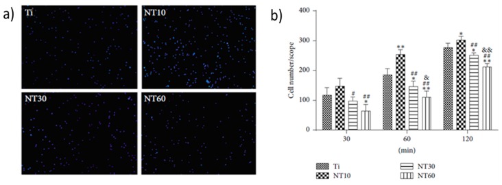
Figure 6.
Fluorescence images of initial adherent bone marrow mesenchymal stem cells (BMSCs) stained with DAPI after 1 h (a) and cell numbers measured by counting cells for 0.5 h, 1 h, and 2 h (b). Cell attachment on NT10 (titanium nanotube with 10 nm diameter) was significantly improved compared with control Ti surfaces for 1 h and 2 h. In contrast, cell attachment was inhibited on NT30 (titanium nanotube with 30 nm diameter) and NT60 (titanium nanotube with 60 nm diameter) at each time interval adopted in this study. All data were expressed as the mean ± standard deviation (SD) and ‘p’, the level of significance. p < 0.05 was considered significant, and p < 0.01 was considered highly significant. Here, the mean ± SD N = 3, and * p < 0.05, and ** p < 0.01 compared with the T; # p < 0.05 and ## p < 0.01 compared with the NT10; & p < 0.05 and && p < 0.01 compared with NT30. Reproduced with permission from [124].

