
Figure 2. TAI exposure causes up-regulation of miR-34a-5p and parallel down-regulation of Bdnf.
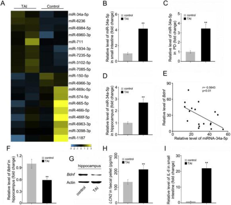
(A) Microarray analysis was performed using small intestine tissues from mice with or without TAI. (B–D) The levels of miR-34a-5p in small intestine (B), PB (C) and hippocampus (D) were assessed by qRT-PCR. (E) The correlation between Bdnf mRNA and miR-34a-5p level was examined by qRT-PCR in 18 cases of hippocampus tissues from TAI exposed mice (P < 0.01; Pearson correlation coefficient, r = − 0.5643). (F, G) The mRNA and protein levels of Bdnf were assessed by qRT-PCR and Western blotting in hippocampus from mice with or without TAI. (H) The level of faecal LCN2 was measured by ELISA from mice with or without TAI. (I) The mRNA level of IL-6 in small intestine tissues was examined by qRT-PCR from mice with or without TAI. Statistically significant differences are indicated: **P < 0.01; Student t-test, n = 18 mice/group.