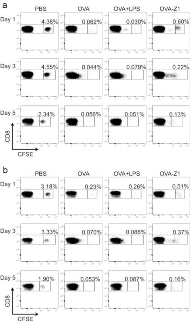
Figure 8.

Immunization with peptide vaccines resulted in CD8+ T cell proliferation in vivo. The frequency of unproliferated CFSE labeled CD8+ cells was gated out in each plot as the evidence of proliferation of CFSE+ cells upon immunizations compared to PBS controls. PBS controls showed no CFSE dilution. OVA–Z1 treated groups showed delayed CD8+ T cell proliferation compared to OVA and OVA+LPS treated groups. Flow cytometry dot plots displaying the CFSE dilution of CD8+ cells from IGLN (a) and spleens (b). The frequency of unproliferated CFSE labeled CD8+ cells was gated out in each plot as the evidence of proliferation of CFSE+ cells upon immunizations compared to PBS controls. All PBS controls showed no CFSE dilution.
