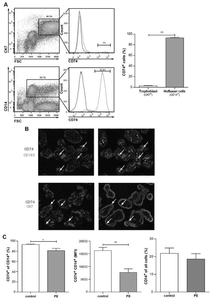
Figure 2. CD74 is highly expressed in placental macrophages (Hofbauer cells) and downregulated in preeclamptic Hofbauer cells.
A) By flow cytometry; representative CK7-positive gating (trophoblast marker) and CD14-positve gating (macrophage marker) is shown for whole placenta cell population (left panel) of healthy controls. Solely 2.6±0.8 % of trophoblasts (CK7-positive) vs. 93.1±1.1 % of Hofbauer cells (CD14-positive) were positive for CD74 staining (right panel) (n=6; p<0.01; Mann Whitney test). B) Immunostaining showed that CD74 (red) is co-localized with CD163 (green) (Hofbauer cells; open arrows; upper panel) but not with CK7 (green). C) Flow cytometry on whole placenta cell population revealed that CD74 was less present in CD14-positive cells of placentas from late preeclamptic women (PE) compared to healthy women (control) (left panel). The mean fluorescent intensity (MFI) of CD74-CD14-positive cells was lower in PE vs. control (middle panel). Percentage of CD14-positive cells in all placental cells was not changed in PE (n=10) vs. control (n=6) (right panel) (*p<0.05, **p<0.01; Mann Whitney test).

