
Figure 2.
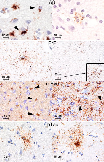
Various type of PAG. Amyloid‐β (Aβ; left: frontal cortex; right: frontal white matter; antibody used: anti‐Aβ, clone 6F/3D directed against amino acids 8–17 of the peptide, Dako, Glostrup, Denmark, 1:100), prion protein (PrP: temporal cortex; antibody used: anti‐PrP 12F10, Cayman Chemical, Ann Arbor, MI, USA, 1:2000), and α‐synuclein (left: striatum; right: temporal cortex; antibody used: anti‐α‐synuclein 5G4, Roboscreen, Leipzig, Germany, 1:4000) PAG showing coarse and fine granules of immunoreactivity: compare with granular/fuzzy astrocytes of ARTAG (temporal cortex; antibody used: anti‐tau AT8 pS202/pT205, Pierce Biotechnology, Rockford, IL, USA, 1:200).