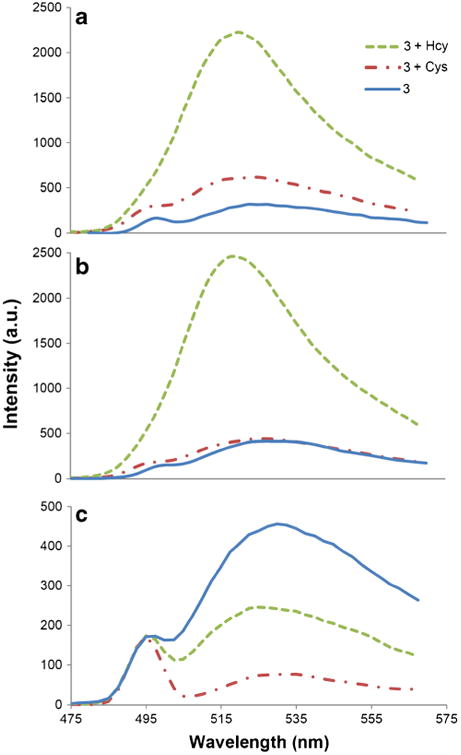
Fig. 5.

Fluorescence spectra of 3 due to Cys and Hcy in buffer after 2 h. (a) At pH 5.0 in phosphate buffer, (b) at pH 6.0 in phosphate buffer, (c) at pH 9.5 in carbonate buffer. Solutions are composed of 3 (4 μM) and analyte (1 mM) in 100 mM buffer solution: DMSO(99:1) at 20 °C, λex = 495 nm
