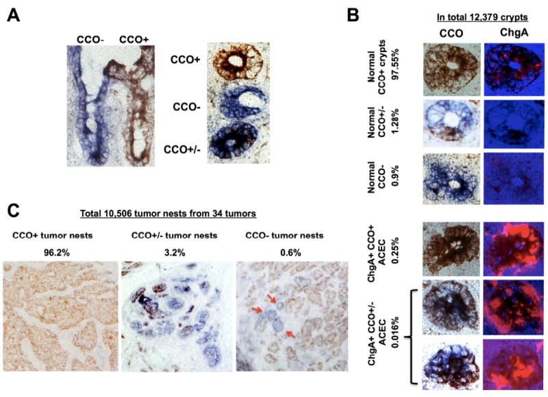
Figure 6.

Frequency and distribution of cytochrome c oxidase (CCO) enzyme deficiency in human normal ileal mucosa and the SI-NETs. (A) Left: CCO enzyme histochemistry of representative frozen sagittal (left) and cross-sections (right) of normal ileal mucosa from a patient with familial SI-NET (F13) illustrating the presence of neighboring CCO- (blue) and CCO+ (brown) and CCO+/- (mixture) crypt-villus structures. (B) Representative images of five groups of crypts categorized on the basis of CCO histochemistry and ChgA immunofluorescence in an ileal segment from a patient with familial SI-NET (F13). The frequency of each category is given as a percent of the 12,379 crypts examined. (C) Representative images of three groups of tumor cell nests categorized on the basis of CCO histochemistry of ileal tumors from a patient with familial SI-NET (F13). The frequency of each category is given as a percent of the 10,506 tumor nests examined from 34 tumors. Tumor nests appear as either CCO+ (brown, left), CCO+/- (mixture of brown and blue, middle) or pure CCO- (blue, right). While the majority of tumor nests are CCO+ (brown), three notable CCO- (blue) tumor nests (red arrows) are present in this tumor.
